Tungsten »
PDB 8c0z-9beo »
8j83 »
Tungsten in PDB 8j83: Crystal Structure of Formate Dehydrogenase From Methylorubrum Extorquens AM1
Enzymatic activity of Crystal Structure of Formate Dehydrogenase From Methylorubrum Extorquens AM1
All present enzymatic activity of Crystal Structure of Formate Dehydrogenase From Methylorubrum Extorquens AM1:
1.17.1.9; 1.2.1.2;
1.17.1.9; 1.2.1.2;
Protein crystallography data
The structure of Crystal Structure of Formate Dehydrogenase From Methylorubrum Extorquens AM1, PDB code: 8j83
was solved by
A.Kobayashi,
M.Taketa,
K.Sowa,
K.Kano,
Y.Higuchi,
H.Ogata,
with X-Ray Crystallography technique. A brief refinement statistics is given in the table below:
| Resolution Low / High (Å) | 47.48 / 2.40 |
| Space group | C 1 2 1 |
| Cell size a, b, c (Å), α, β, γ (°) | 232.86, 73.95, 95.74, 90, 106.72, 90 |
| R / Rfree (%) | 21.9 / 25.9 |
Other elements in 8j83:
The structure of Crystal Structure of Formate Dehydrogenase From Methylorubrum Extorquens AM1 also contains other interesting chemical elements:
| Iron | (Fe) | 20 atoms |
Tungsten Binding Sites:
The binding sites of Tungsten atom in the Crystal Structure of Formate Dehydrogenase From Methylorubrum Extorquens AM1
(pdb code 8j83). This binding sites where shown within
5.0 Angstroms radius around Tungsten atom.
In total only one binding site of Tungsten was determined in the Crystal Structure of Formate Dehydrogenase From Methylorubrum Extorquens AM1, PDB code: 8j83:
In total only one binding site of Tungsten was determined in the Crystal Structure of Formate Dehydrogenase From Methylorubrum Extorquens AM1, PDB code: 8j83:
Tungsten binding site 1 out of 1 in 8j83
Go back to
Tungsten binding site 1 out
of 1 in the Crystal Structure of Formate Dehydrogenase From Methylorubrum Extorquens AM1
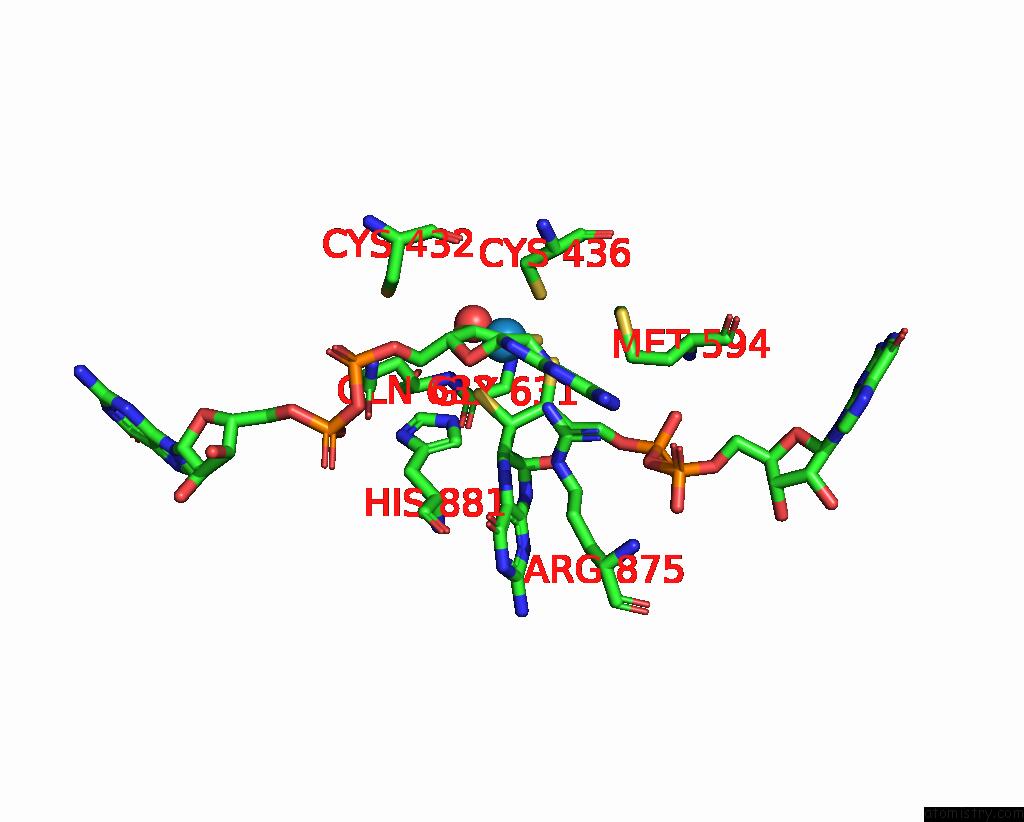
Mono view
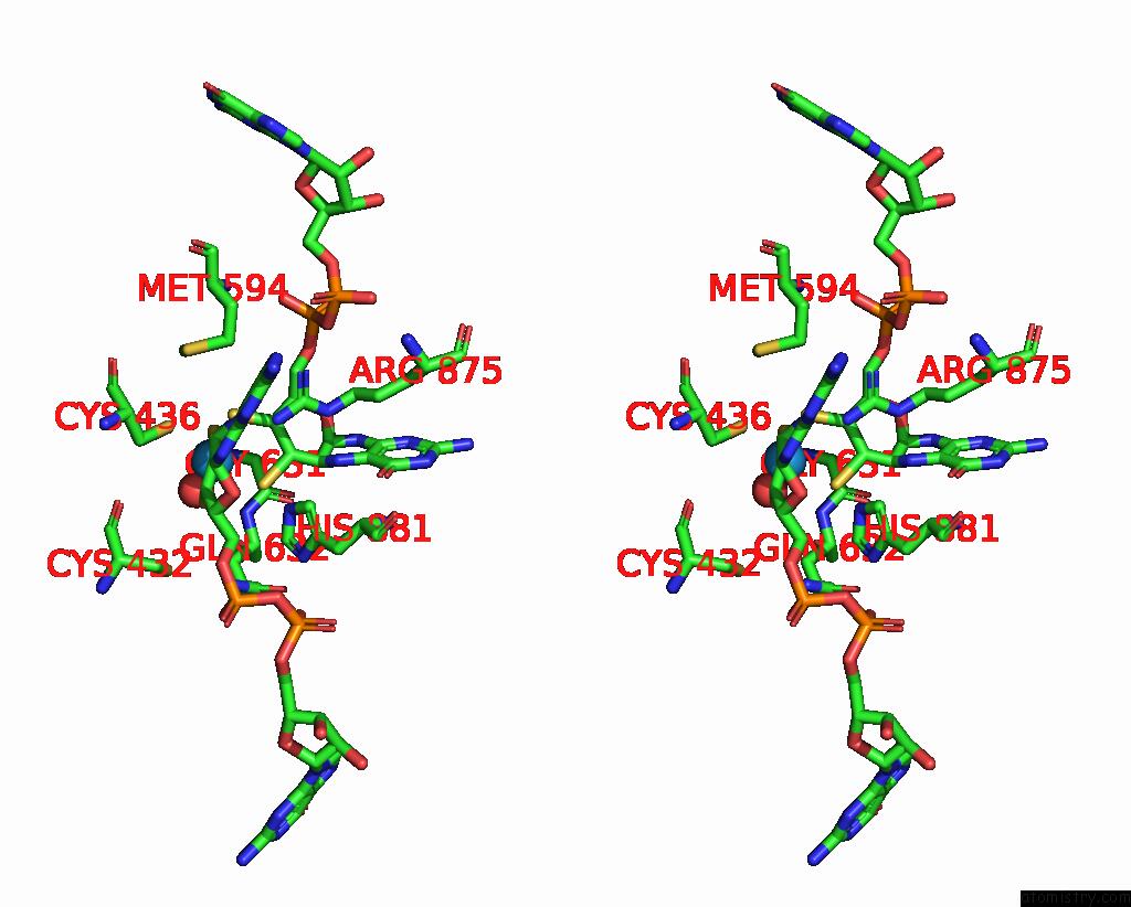
Stereo pair view
Mono view
Stereo pair view
A full contact list of Tungsten with other atoms in the W binding
site number 1 of Crystal Structure of Formate Dehydrogenase From Methylorubrum Extorquens AM1 within 5.0Å range:
|
Reference:
A.Kobayashi,
M.Taketa,
K.Sowa,
K.Kano,
Y.Higuchi,
H.Ogata.
Structure and Function Relationship of Formate Dehydrogenases: An Overview of Recent Progress. Iucrj V. 10 544 2023.
ISSN: ESSN 2052-2525
PubMed: 37668215
DOI: 10.1107/S2052252523006437
Page generated: Sat Oct 12 15:56:55 2024
ISSN: ESSN 2052-2525
PubMed: 37668215
DOI: 10.1107/S2052252523006437
Last articles
Zn in 9J0NZn in 9J0O
Zn in 9J0P
Zn in 9FJX
Zn in 9EKB
Zn in 9C0F
Zn in 9CAH
Zn in 9CH0
Zn in 9CH3
Zn in 9CH1