Tungsten »
PDB 6sdr-8bql »
8bql »
Tungsten in PDB 8bql: W-Formate Dehydrogenase From Desulfovibrio Vulgaris - Co-Crystallized with Formate and Reoxidized By Exposure to Air For 12 Min
Protein crystallography data
The structure of W-Formate Dehydrogenase From Desulfovibrio Vulgaris - Co-Crystallized with Formate and Reoxidized By Exposure to Air For 12 Min, PDB code: 8bql
was solved by
G.Vilela-Alves,
C.Mota,
A.R.Oliveira,
R.R.Manuel,
I.C.Pereira,
M.J.Romao,
with X-Ray Crystallography technique. A brief refinement statistics is given in the table below:
| Resolution Low / High (Å) | 97.00 / 1.91 |
| Space group | P 21 21 21 |
| Cell size a, b, c (Å), α, β, γ (°) | 64.8, 127.594, 148.622, 90, 90, 90 |
| R / Rfree (%) | 18.8 / 22.5 |
Other elements in 8bql:
The structure of W-Formate Dehydrogenase From Desulfovibrio Vulgaris - Co-Crystallized with Formate and Reoxidized By Exposure to Air For 12 Min also contains other interesting chemical elements:
| Iron | (Fe) | 16 atoms |
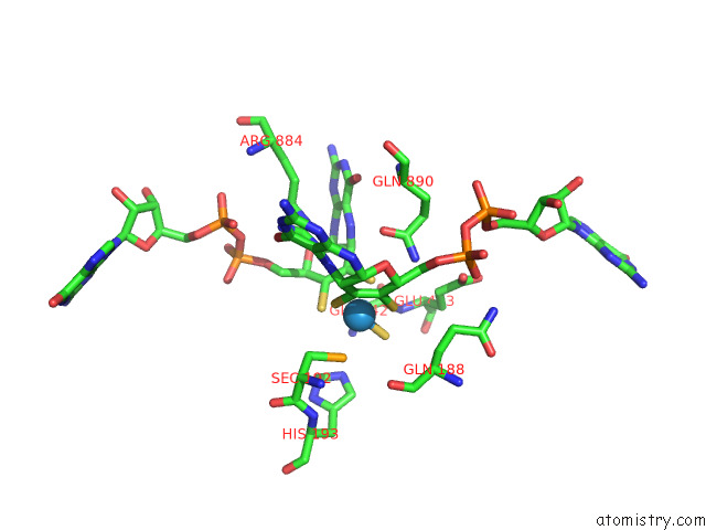
Tungsten Binding Sites:
The binding sites of Tungsten atom in the W-Formate Dehydrogenase From Desulfovibrio Vulgaris - Co-Crystallized with Formate and Reoxidized By Exposure to Air For 12 Min
(pdb code 8bql). This binding sites where shown within
5.0 Angstroms radius around Tungsten atom.
In total only one binding site of Tungsten was determined in the W-Formate Dehydrogenase From Desulfovibrio Vulgaris - Co-Crystallized with Formate and Reoxidized By Exposure to Air For 12 Min, PDB code: 8bql:
In total only one binding site of Tungsten was determined in the W-Formate Dehydrogenase From Desulfovibrio Vulgaris - Co-Crystallized with Formate and Reoxidized By Exposure to Air For 12 Min, PDB code: 8bql:
Tungsten binding site 1 out of 1 in 8bql
Go back to
Tungsten binding site 1 out
of 1 in the W-Formate Dehydrogenase From Desulfovibrio Vulgaris - Co-Crystallized with Formate and Reoxidized By Exposure to Air For 12 Min
Mono view
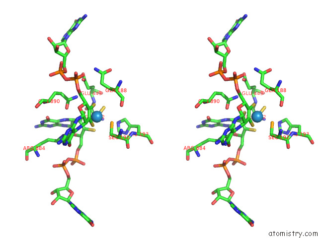
Stereo pair view
Mono view
Stereo pair view
A full contact list of Tungsten with other atoms in the W binding
site number 1 of W-Formate Dehydrogenase From Desulfovibrio Vulgaris - Co-Crystallized with Formate and Reoxidized By Exposure to Air For 12 Min within 5.0Å range:
|
Reference:
G.Vilela-Alves,
R.R.Manuel,
A.R.Oliveira,
I.C.Pereira,
M.J.Romao,
C.Mota.
Tracking W-Formate Dehydrogenase Structural Changes During Catalysis and Enzyme Reoxidation. Int J Mol Sci V. 24 2022.
ISSN: ESSN 1422-0067
PubMed: 36613918
DOI: 10.3390/IJMS24010476
Page generated: Sat Oct 12 15:46:42 2024
ISSN: ESSN 1422-0067
PubMed: 36613918
DOI: 10.3390/IJMS24010476
Last articles
Zn in 9J0NZn in 9J0O
Zn in 9J0P
Zn in 9FJX
Zn in 9EKB
Zn in 9C0F
Zn in 9CAH
Zn in 9CH0
Zn in 9CH3
Zn in 9CH1