Tungsten »
PDB 6sdr-8bql »
8bqg »
Tungsten in PDB 8bqg: W-Formate Dehydrogenase From Desulfovibrio Vulgaris - Soaking with Formate 1 Min
Protein crystallography data
The structure of W-Formate Dehydrogenase From Desulfovibrio Vulgaris - Soaking with Formate 1 Min, PDB code: 8bqg
was solved by
G.Vilela-Alves,
C.Mota,
A.R.Oliveira,
R.R.Manuel,
I.C.Pereira,
M.J.Romao,
with X-Ray Crystallography technique. A brief refinement statistics is given in the table below:
| Resolution Low / High (Å) | 94.93 / 1.95 |
| Space group | P 21 21 21 |
| Cell size a, b, c (Å), α, β, γ (°) | 64.537, 122.778, 148.985, 90, 90, 90 |
| R / Rfree (%) | 19.3 / 24.3 |
Other elements in 8bqg:
The structure of W-Formate Dehydrogenase From Desulfovibrio Vulgaris - Soaking with Formate 1 Min also contains other interesting chemical elements:
| Iron | (Fe) | 16 atoms |
Tungsten Binding Sites:
The binding sites of Tungsten atom in the W-Formate Dehydrogenase From Desulfovibrio Vulgaris - Soaking with Formate 1 Min
(pdb code 8bqg). This binding sites where shown within
5.0 Angstroms radius around Tungsten atom.
In total only one binding site of Tungsten was determined in the W-Formate Dehydrogenase From Desulfovibrio Vulgaris - Soaking with Formate 1 Min, PDB code: 8bqg:
In total only one binding site of Tungsten was determined in the W-Formate Dehydrogenase From Desulfovibrio Vulgaris - Soaking with Formate 1 Min, PDB code: 8bqg:
Tungsten binding site 1 out of 1 in 8bqg
Go back to
Tungsten binding site 1 out
of 1 in the W-Formate Dehydrogenase From Desulfovibrio Vulgaris - Soaking with Formate 1 Min
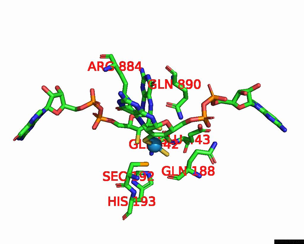
Mono view
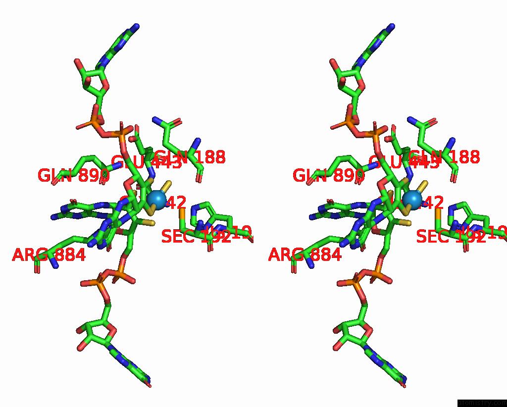
Stereo pair view
Mono view
Stereo pair view
A full contact list of Tungsten with other atoms in the W binding
site number 1 of W-Formate Dehydrogenase From Desulfovibrio Vulgaris - Soaking with Formate 1 Min within 5.0Å range:
|
Reference:
G.Vilela-Alves,
R.R.Manuel,
A.R.Oliveira,
I.C.Pereira,
M.J.Romao,
C.Mota.
Tracking W-Formate Dehydrogenase Structural Changes During Catalysis and Enzyme Reoxidation. Int J Mol Sci V. 24 2022.
ISSN: ESSN 1422-0067
PubMed: 36613918
DOI: 10.3390/IJMS24010476
Page generated: Sat Oct 12 15:43:07 2024
ISSN: ESSN 1422-0067
PubMed: 36613918
DOI: 10.3390/IJMS24010476
Last articles
Ca in 2P5VCa in 2P5R
Ca in 2P69
Ca in 2P5W
Ca in 2P1R
Ca in 2P3U
Ca in 2P4P
Ca in 2P3T
Ca in 2P37
Ca in 2P34