Tungsten »
PDB 4z3x-6rvg »
5jki »
Tungsten in PDB 5jki: Crystal Structure of the First Transmembrane PAP2 Type Phosphatidylglycerolphosphate Phosphatase From Bacillus Subtilis
Protein crystallography data
The structure of Crystal Structure of the First Transmembrane PAP2 Type Phosphatidylglycerolphosphate Phosphatase From Bacillus Subtilis, PDB code: 5jki
was solved by
M.El Ghachi,
N.Howe,
A.Lampion,
F.Delbrassine,
L.Vogeley,
M.Caffrey,
E.Sauvage,
R.Auger,
A.Guiseppe,
S.Roure,
S.Perlier,
D.Mengin-Lecreulx,
M.Foglino,
T.Touze,
with X-Ray Crystallography technique. A brief refinement statistics is given in the table below:
| Resolution Low / High (Å) | 38.48 / 2.25 |
| Space group | I 2 2 2 |
| Cell size a, b, c (Å), α, β, γ (°) | 70.066, 76.961, 99.629, 90.00, 90.00, 90.00 |
| R / Rfree (%) | 20.1 / 23.1 |
Tungsten Binding Sites:
The binding sites of Tungsten atom in the Crystal Structure of the First Transmembrane PAP2 Type Phosphatidylglycerolphosphate Phosphatase From Bacillus Subtilis
(pdb code 5jki). This binding sites where shown within
5.0 Angstroms radius around Tungsten atom.
In total only one binding site of Tungsten was determined in the Crystal Structure of the First Transmembrane PAP2 Type Phosphatidylglycerolphosphate Phosphatase From Bacillus Subtilis, PDB code: 5jki:
In total only one binding site of Tungsten was determined in the Crystal Structure of the First Transmembrane PAP2 Type Phosphatidylglycerolphosphate Phosphatase From Bacillus Subtilis, PDB code: 5jki:
Tungsten binding site 1 out of 1 in 5jki
Go back to
Tungsten binding site 1 out
of 1 in the Crystal Structure of the First Transmembrane PAP2 Type Phosphatidylglycerolphosphate Phosphatase From Bacillus Subtilis
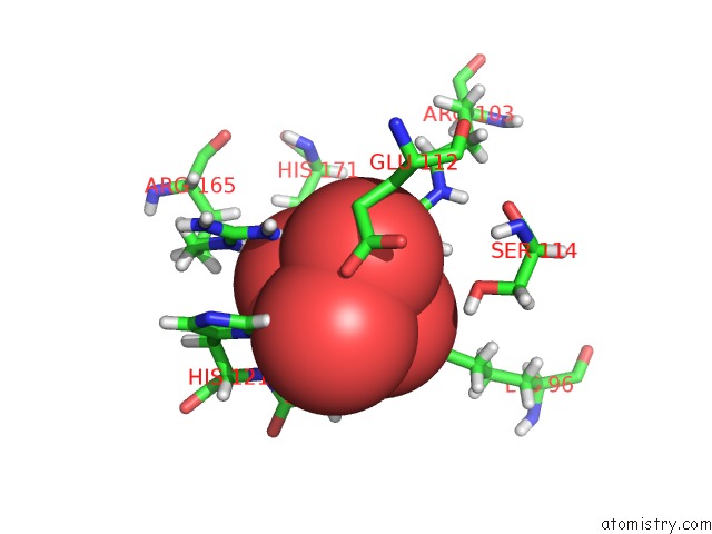
Mono view
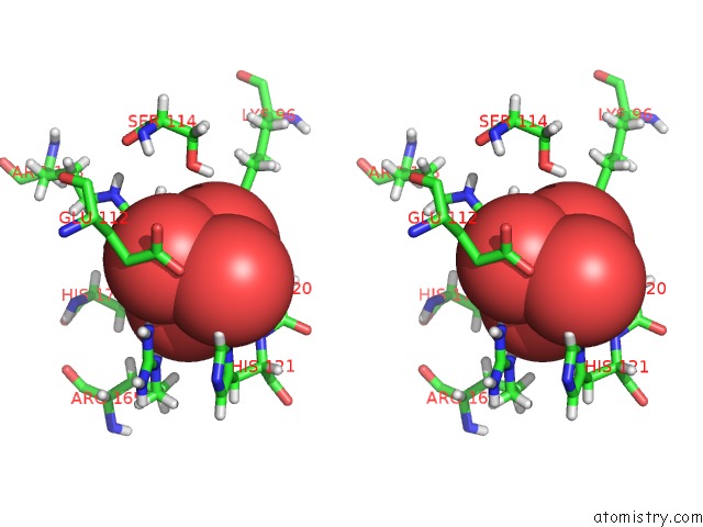
Stereo pair view
Mono view
Stereo pair view
A full contact list of Tungsten with other atoms in the W binding
site number 1 of Crystal Structure of the First Transmembrane PAP2 Type Phosphatidylglycerolphosphate Phosphatase From Bacillus Subtilis within 5.0Å range:
|
Reference:
M.E.Ghachi,
N.Howe,
R.Auger,
A.Lambion,
A.Guiseppi,
F.Delbrassine,
G.Manat,
S.Roure,
S.Peslier,
E.Sauvage,
L.Vogeley,
J.C.Rengifo-Gonzalez,
P.Charlier,
D.Mengin-Lecreulx,
M.Foglino,
T.Touze,
M.Caffrey,
F.Kerff.
Crystal Structure and Biochemical Characterization of the Transmembrane PAP2 Type Phosphatidylglycerol Phosphate Phosphatase From Bacillus Subtilis. Cell. Mol. Life Sci. V. 74 2319 2017.
ISSN: ESSN 1420-9071
PubMed: 28168443
DOI: 10.1007/S00018-017-2464-6
Page generated: Sat Oct 12 04:50:31 2024
ISSN: ESSN 1420-9071
PubMed: 28168443
DOI: 10.1007/S00018-017-2464-6
Last articles
Fe in 2YXOFe in 2YRS
Fe in 2YXC
Fe in 2YNM
Fe in 2YVJ
Fe in 2YP1
Fe in 2YU2
Fe in 2YU1
Fe in 2YQB
Fe in 2YOO