Tungsten »
PDB 2hhl-4z3w »
3et5 »
Tungsten in PDB 3et5: Structure of Recombinant Haemophilus Influenzae E(P4) Acid Phosphatase Complexed with Tungstate
Enzymatic activity of Structure of Recombinant Haemophilus Influenzae E(P4) Acid Phosphatase Complexed with Tungstate
All present enzymatic activity of Structure of Recombinant Haemophilus Influenzae E(P4) Acid Phosphatase Complexed with Tungstate:
3.1.3.2;
3.1.3.2;
Protein crystallography data
The structure of Structure of Recombinant Haemophilus Influenzae E(P4) Acid Phosphatase Complexed with Tungstate, PDB code: 3et5
was solved by
J.J.Tanner,
with X-Ray Crystallography technique. A brief refinement statistics is given in the table below:
| Resolution Low / High (Å) | 42.30 / 2.00 |
| Space group | P 42 21 2 |
| Cell size a, b, c (Å), α, β, γ (°) | 65.848, 65.848, 101.893, 90.00, 90.00, 90.00 |
| R / Rfree (%) | 18.2 / 22.5 |
Other elements in 3et5:
The structure of Structure of Recombinant Haemophilus Influenzae E(P4) Acid Phosphatase Complexed with Tungstate also contains other interesting chemical elements:
| Magnesium | (Mg) | 1 atom |
Tungsten Binding Sites:
The binding sites of Tungsten atom in the Structure of Recombinant Haemophilus Influenzae E(P4) Acid Phosphatase Complexed with Tungstate
(pdb code 3et5). This binding sites where shown within
5.0 Angstroms radius around Tungsten atom.
In total only one binding site of Tungsten was determined in the Structure of Recombinant Haemophilus Influenzae E(P4) Acid Phosphatase Complexed with Tungstate, PDB code: 3et5:
In total only one binding site of Tungsten was determined in the Structure of Recombinant Haemophilus Influenzae E(P4) Acid Phosphatase Complexed with Tungstate, PDB code: 3et5:
Tungsten binding site 1 out of 1 in 3et5
Go back to
Tungsten binding site 1 out
of 1 in the Structure of Recombinant Haemophilus Influenzae E(P4) Acid Phosphatase Complexed with Tungstate

Mono view
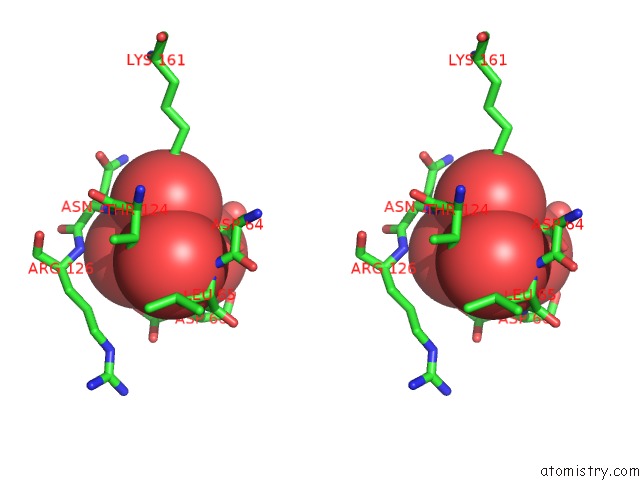
Stereo pair view
Mono view
Stereo pair view
A full contact list of Tungsten with other atoms in the W binding
site number 1 of Structure of Recombinant Haemophilus Influenzae E(P4) Acid Phosphatase Complexed with Tungstate within 5.0Å range:
|
Reference:
R.L.Felts,
Z.Ou,
T.J.Reilly,
J.J.Tanner.
Structure of Recombinant Haemophilus Influenzae E (P4) Acid Phosphatase Reveals A New Member of the Haloacid Dehalogenase Superfamily. Biochemistry V. 46 11110 2007.
ISSN: ISSN 0006-2960
PubMed: 17824671
DOI: 10.1021/BI701016M
Page generated: Sat Oct 12 04:06:09 2024
ISSN: ISSN 0006-2960
PubMed: 17824671
DOI: 10.1021/BI701016M
Last articles
F in 7PK3F in 7PJC
F in 7PJ2
F in 7PHN
F in 7PG6
F in 7PHJ
F in 7PAV
F in 7PH1
F in 7PCD
F in 7P80